
Diabetes is one of the leading causes of blindness in adults in the United States — but here is the good news: most diabetes-related vision loss is preventable. The key is catching problems early with a regular diabetic eye exam. At Eye Medics Optometry in Fayetteville, NC, we provide comprehensive diabetic eye care for patients throughout Cumberland County, including Hope Mills, Spring Lake, Raeford, and the Fort Liberty community.
According to the Centers for Disease Control and Prevention (CDC), about 60% of Americans with diabetes do not get the recommended annual eye exam — even though early detection can prevent most diabetes-related vision loss. Do not be part of that statistic.
What Is a Diabetic Eye Exam?
A diabetic eye exam is a comprehensive eye evaluation specifically designed for people with type 1 or type 2 diabetes. It goes beyond checking your vision prescription. The exam focuses on the health of the blood vessels in your retina — the light-sensitive tissue at the back of your eye — because high blood sugar over time can damage those vessels and lead to serious vision problems.
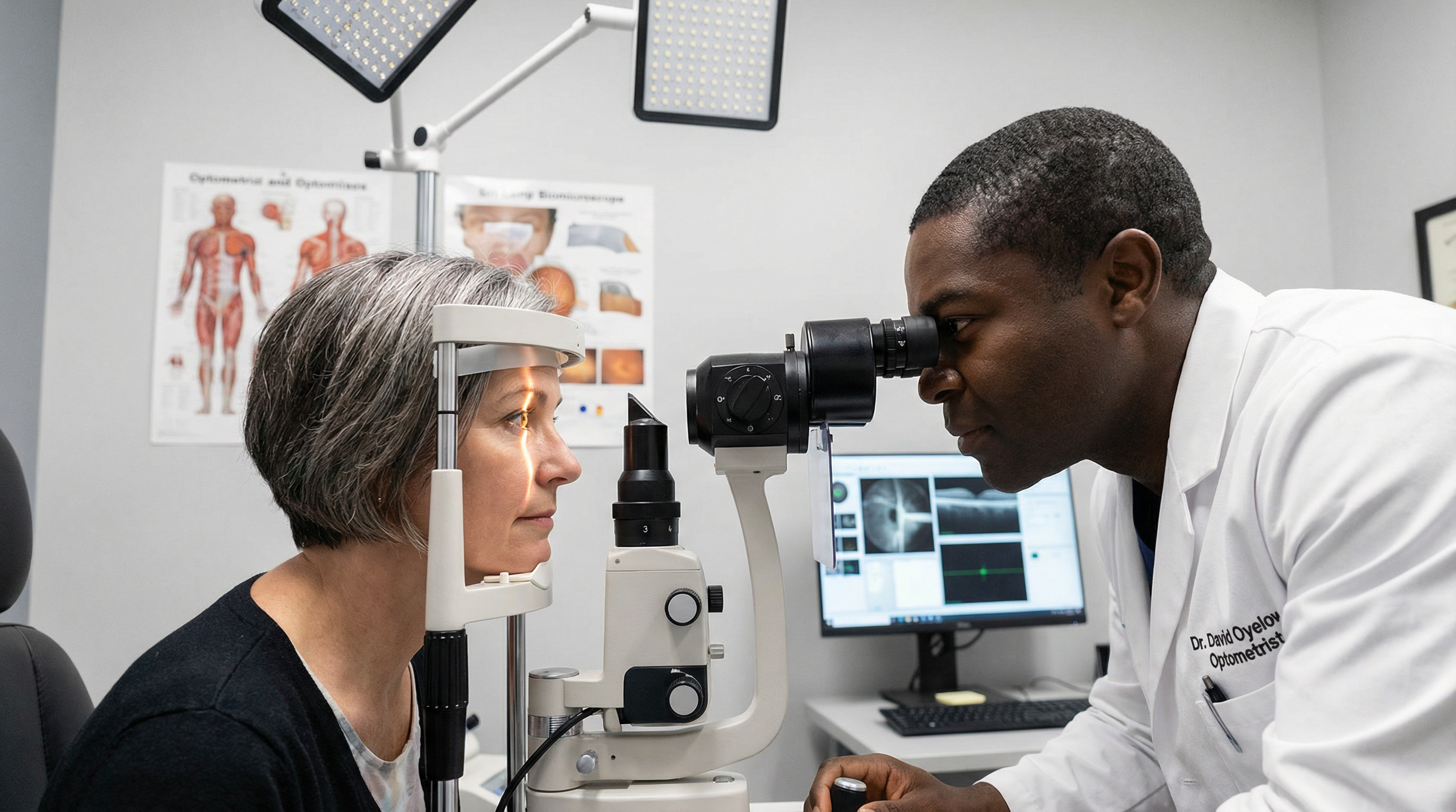
The most important part of a diabetic eye exam is the dilated fundus exam, where we use special eye drops to widen your pupils so we can see the full retina. We also use advanced imaging technology — including optical coherence tomography (OCT) and retinal photography — to capture detailed images of your retina and optic nerve.
Why Diabetic Eye Exams Are So Important
High blood sugar damages the tiny blood vessels throughout your body — including the ones in your eyes. Over time, this can lead to diabetic retinopathy, diabetic macular edema, glaucoma, and cataracts. The dangerous thing about these conditions is that they often cause no symptoms in the early stages.
Risk of Severe Vision Loss: Exams vs. No Exams
Early detection with regular exams can reduce the risk of severe vision loss from diabetic eye disease by up to 95%. (Source: American Diabetes Association)
The American Diabetes Association states that early detection and timely treatment can reduce the risk of severe vision loss from diabetic eye disease by 95%. That is nearly complete prevention when you stay on top of your annual exams.
Diabetic retinopathy affects approximately 30% of people with diabetes. With over 37 million Americans living with diabetes, that means millions of people are at risk of losing their sight — many of them without knowing it.
How a Diabetic Eye Exam Differs from a Regular Eye Exam
A standard eye exam checks your vision, eye pressure, and general eye health. A diabetic eye exam includes all of that — plus a much more detailed look at the structures most affected by diabetes:
Standard Eye Exam Includes:
- Visual acuity test (reading chart)
- Eye pressure check (glaucoma screening)
- Basic retinal view
- Prescription check
Diabetic Eye Exam Also Includes:
- Pupil dilation for full retinal view
- Retinal photography and OCT imaging
- Detailed optic nerve assessment
- Screening for microaneurysms and hemorrhages
- Macular edema evaluation
- Coordination with your diabetes care team
What to Expect at Your Diabetic Eye Exam
Your appointment at Eye Medics will typically last 60 to 90 minutes. Here is what happens from start to finish:
- 1
Medical History Review
We will ask about your type of diabetes, how long you have had it, your most recent A1C levels, blood pressure, and any medications you take. This helps us understand your overall risk level.
- 2
Visual Acuity Test
You will read letters on a chart to check how clearly you can see at different distances.
- 3
Eye Pressure Measurement (Tonometry)
We measure the pressure inside your eyes to screen for glaucoma, which is more common in people with diabetes.
- 4
Pupil Dilation
We apply dilating eye drops that take about 15 to 20 minutes to work. Your vision will be blurry and light-sensitive for a few hours after the exam.
- 5
Dilated Fundus Exam
Using a special lens and bright light, Dr. Singletary examines your retina, macula, optic nerve, and blood vessels in detail.
- 6
Retinal Imaging and OCT
We take high-resolution photographs and cross-sectional scans of your retina to document your baseline and detect any changes over time.
- 7
Results and Next Steps
We will explain what we found, answer your questions, and coordinate with your primary care doctor or endocrinologist if needed.
Eye Conditions Linked to Diabetes
Diabetes can affect your eyes in several ways. Understanding these conditions helps you know what we are looking for and why early detection matters so much.
Diabetic Retinopathy
The most common diabetic eye disease. High blood sugar damages the blood vessels in the retina, causing them to leak, swell, or grow new abnormal vessels. There are four stages, from mild to proliferative (see chart below).
Diabetic Macular Edema (DME)
Fluid builds up in the macula — the central part of the retina responsible for sharp, detailed vision. DME can occur at any stage of diabetic retinopathy and is a leading cause of vision loss in people with diabetes.
Glaucoma
People with diabetes are twice as likely to develop glaucoma — a condition where increased eye pressure damages the optic nerve. We screen for glaucoma at every diabetic eye exam.
Cataracts
Diabetes causes cataracts to develop earlier and progress faster than in people without diabetes. Cataracts cloud the natural lens of the eye and cause blurry, hazy vision.
| Stage | What Is Happening | Symptoms | Recommended Action |
|---|---|---|---|
| Mild NPDR | Small areas of swelling (microaneurysms) in retinal blood vessels | Usually none | Annual monitoring |
| Moderate NPDR | Blood vessels that feed the retina begin to swell and lose ability to transport blood | Possible mild blurring | More frequent exams |
| Severe NPDR | Many blood vessels are blocked, depriving areas of the retina of blood supply | Blurred or patchy vision | Prompt treatment |
| Proliferative DR (PDR) | New, fragile blood vessels grow on the retina — these can bleed and cause serious vision loss | Floaters, vision loss | Urgent treatment |
NPDR = Non-Proliferative Diabetic Retinopathy. Source: American Diabetes Association, National Eye Institute.
How Often Should You Get a Diabetic Eye Exam?
The frequency of your diabetic eye exams depends on your situation:
| Your Situation | Recommended Frequency |
|---|---|
| Newly diagnosed with type 1 or type 2 diabetes | Within 5 years of diagnosis (type 1); at diagnosis (type 2) |
| No diabetic eye disease detected | Once a year |
| Mild to moderate diabetic retinopathy | Every 6 to 12 months |
| Severe retinopathy or DME | Every 3 to 6 months |
| Pregnant with diabetes | First trimester, then as recommended |
Source: American Diabetes Association Clinical Standards of Care.
How to Prepare for Your Diabetic Eye Exam
A little preparation goes a long way toward getting the most out of your visit:
Know your A1C and blood sugar levels
Bring your most recent lab results or diabetes management records.
List all medications
Include insulin, oral medications, blood pressure drugs, and any supplements.
Bring sunglasses
Your pupils will be dilated after the exam, making your eyes sensitive to light for several hours.
Arrange a ride
Dilation can blur your vision temporarily. Having a driver is a good idea, especially for your first exam.
Know your symptoms
Note any recent changes in vision — blurring, floaters, flashes of light, or dark spots.
Bring your insurance card
Medicare and most insurance plans cover annual diabetic eye exams. We will verify your benefits.
Frequently Asked Questions
A diabetic eye exam includes pupil dilation and a detailed examination of the retina and optic nerve — areas that diabetes directly damages. A standard eye exam checks your vision and eye health but may not include the same level of retinal screening that people with diabetes need.
Schedule Your Diabetic Eye Exam Today
Serving patients with diabetes throughout Fayetteville, Hope Mills, Spring Lake, Raeford, and Fort Liberty. Do not wait for symptoms — protect your vision now.
Book a Diabetic Eye Exam